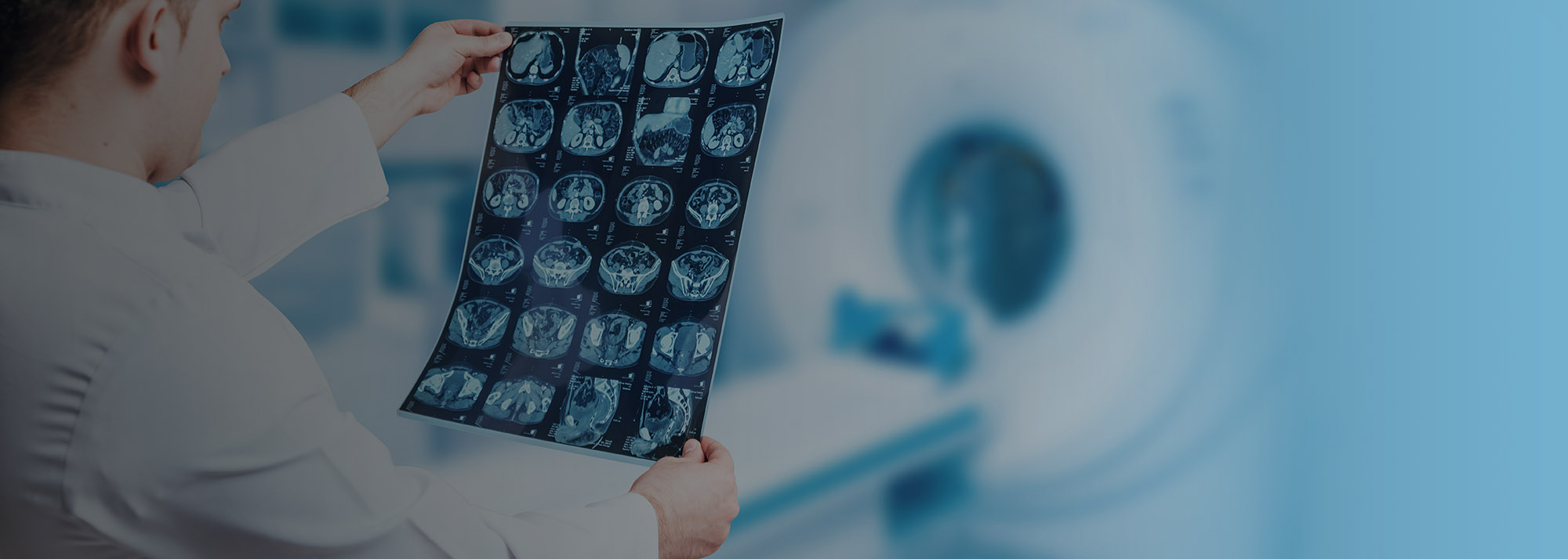
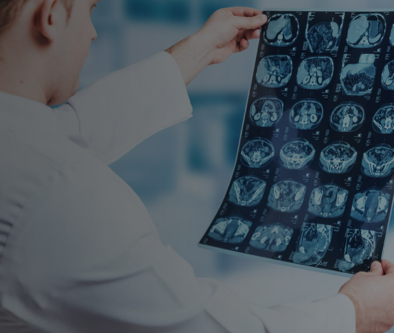
One of the most comprehensive facilities for diagnostic and therapeutic imaging (including Nuclear Medicine), the Clinical Imaging unit at Aster Medcity is equipped with state-of-the-art technology including 3.0 Tesla Wide Bore MRI, 256 Slice Philips iCT scanner, PET CT with Time of Flight technology, SPECT, EPIQ, Image Guided Biopsies, Fluoroscopy facility, DEXA for bone density measurement, Digital Mammogram with Tomosynthesis and vacuum assisted biopsy, Flat Panel Bi Plane Vascular Hybrid Cathlab and Low Radiation Clarity Cath Lab.
The expert team of doctors here comprise Radiologists and Interventional Radiologists with extensive international training and clinical expertise. Multidisciplinary in approach, they provide protocol-based services/ treatment to patients with the help of technicians and other support staff.
Advanced Technology & Facilities
Well equipped with the latest medical equipment, modern technology & infrastructure, Aster Hospital is one of the best hospitals in India.
An advanced Digital Mammography system, it provides high-resolution 3D image results using X-rays for quick detection of even the tiniest calcified lesion that’s in the precancerous stage. The X-ray tubes move in an arc, capturing 11 images in 7 seconds and these images are assembled in the computer to create highly focused 3D image throughout the breast, facilitating easier diagnosis.
The Dexa, utilizing x-ray beams of two different energies, facilitates accurate measurement of bone density and strength with lowest dose of radiation possible, especially for preclinical detection of osteoporosis and preventing complications.
Renders high-resolution radiographs with superior time efficiency. As no repetition and digital enhancement are required, rapid transfer of images for immediate investigation by Physician is made possible through integration with PACS.
Time of Flight PET CT Accurate and early lesion detection of functional abnormalities and low FDG doses using Time of Flight technology
This Catheterisation Lab (Cath Lab), with the help of Allura Clarity system, reduces radiation doses by 70%, offering significant benefit to the patient as well as staff. Used for Interventional Cardiology and Neurology, the low radiation facilitates repeated investigation if necessary and is also safe for children.
The Biplane Hybrid Cath Lab Unit, which works in a sterile operation theatre environment, combines the traditional diagnostic functions of a Cath lab with surgical functions of an operating room. Designed to undertake complex Neuro vascular and peripheral vascular procedures and Hybrid surgical/ endovascular procedures, the Cathlabs here are the first to have the option of ‘Clarity’, an ingenious system that reduces the radiation dose by upto 70%, which is of significant benefit to the patients. The functions of the unit include: Biplane Cath Lab Bi Plane Cath Labs are capable of capturing 3D images in real time, and come with added components like 3D road mapping and post processing to help neurovascular procedures. It can perform interventional procedures in lesser time with lesser usage of contrast, in comparison to single plane. Digital Subtraction Angiography (DSA) A type of Fluoroscopy technique used in interventional radiology to clearly visualise blood vessels in a bony or dense soft tissue environment, images are generated using contrast medium by subtracting a ‘pre-contrast image’. Very helpful in diagnosing Neurological problems, the advantage of DSA is that the interference of bony structures can be eliminated.
3D Rotational Angiography - 3D rotational angiography provides an image of a particular vessel or chamber while the camera rotates around the patient in a predefined arc, enabling viewing of the structure from multiple angles with a single injection of contrast medium, thereby saving the contrast load and its side effects. A faster procedure compared to conventional Angiography, it is beneficial for patients for whom dye load needs to be restricted and those who require abnormal kidney function test. CT Like images With the soft tissue imaging feature that provides enables CT like images, the patient need not to be sent for a CT examination separately. This is a great advantage for neurovascular cases where the mobility of the patient is restricted and timelines are crucial. Interventionist can access CT-like imaging right on the angio system and inturn the soft tissue, bone and other brain structures before, during, or after an interventional procedure. 3D soft tissue imaging supports diagnosis, planning, interventions and treatment follow-up. Hybrid Cath Lab In a hybrid application, the Cath lab doubles as an operating room so that a patient undergoing a cardiac cath procedure can have a surgery if required. Enabling the Doctor to address the patient needs quickly, eliminating the need to schedule an additional surgical procedure. As the hybrid cath lab can turn to any angle as required by the interventionist, and can be placed in the corner of the OT when not in use.
Most sophisticated Colour Doppler Systems with Liver Elastography, fusion and navigation for accurate Biopsy, Electronic 4D Imaging with lightweight probes for Cardiac, Radiology and Obstetric Studies. Also provides superior 4D images of the foetus. Department has many state-of-the-art Ultrasound Scanners available for 4D imaging, non-invasive monitoring of liver parenchymal disease with ARFI (Acoustic Radiation Forced Impulse) imaging, with sophisticated image fusion capability to aid accurate guided procedures.
Ultrasound Machines with multi modality image fusion (like CT and MRI) capability and accurate needle tracking.
Blogs
The source of trustworthy health and medical information. Through this section, we provide research-based health information, and all that is happening in Aster Hospital.
