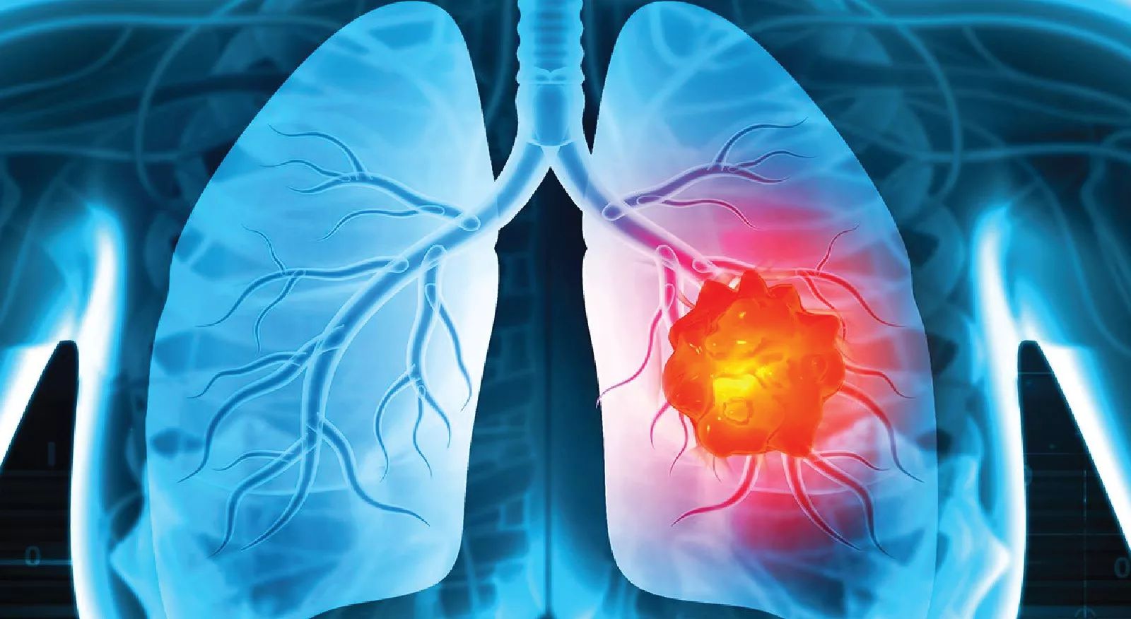
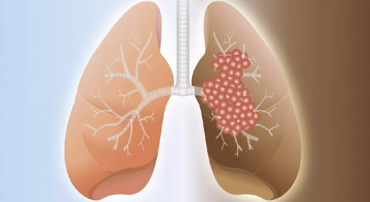
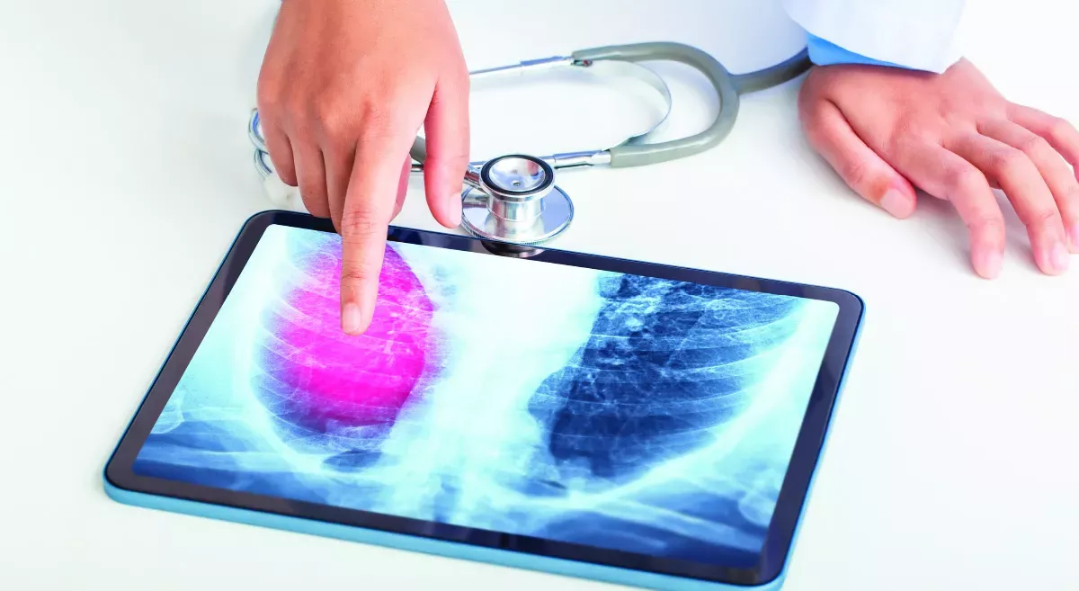
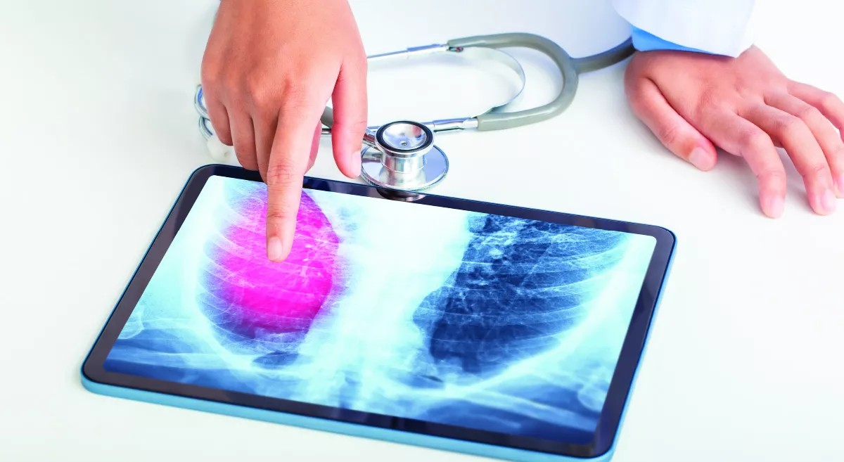
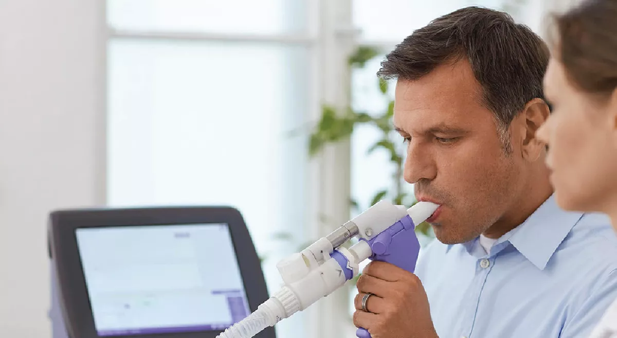
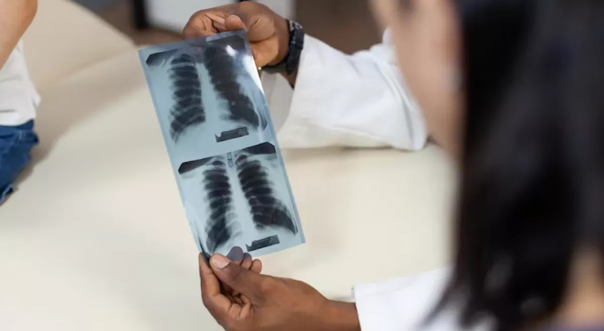
When 52-year-old Ragul ( Name Changed), a non-smoker and fitness enthusiast, started experiencing a persistent cough, he initially thought it was a seasonal allergy. But as weeks turned into months, the cough persisted, and he noticed breathlessness while climbing stairs. Concerned, his wife encouraged him to seek medical advice. His local doctor ordered a chest X-ray, which revealed a suspicious shadow on the right side of his chest. To investigate further, a CT scan of the thorax was performed. The scan showed a localised lesion in the right upper lobe of his lung, raising concerns that something more serious could be happening. This discovery led to a series of advanced diagnostic procedures—bronchoscopy followed by EBUS (Endobronchial Ultrasound)—that ultimately saved Rahul’s life by allowing early, precise diagnosis and timely treatment.
Step 1: The Role of Bronchoscopy in Rahul’s Diagnosis
Given the concerning findings on the CT scan, Rahul’s pulmonologist recommended a bronchoscopy to get a closer look and obtain a biopsy.
What Happened During Bronchoscopy?
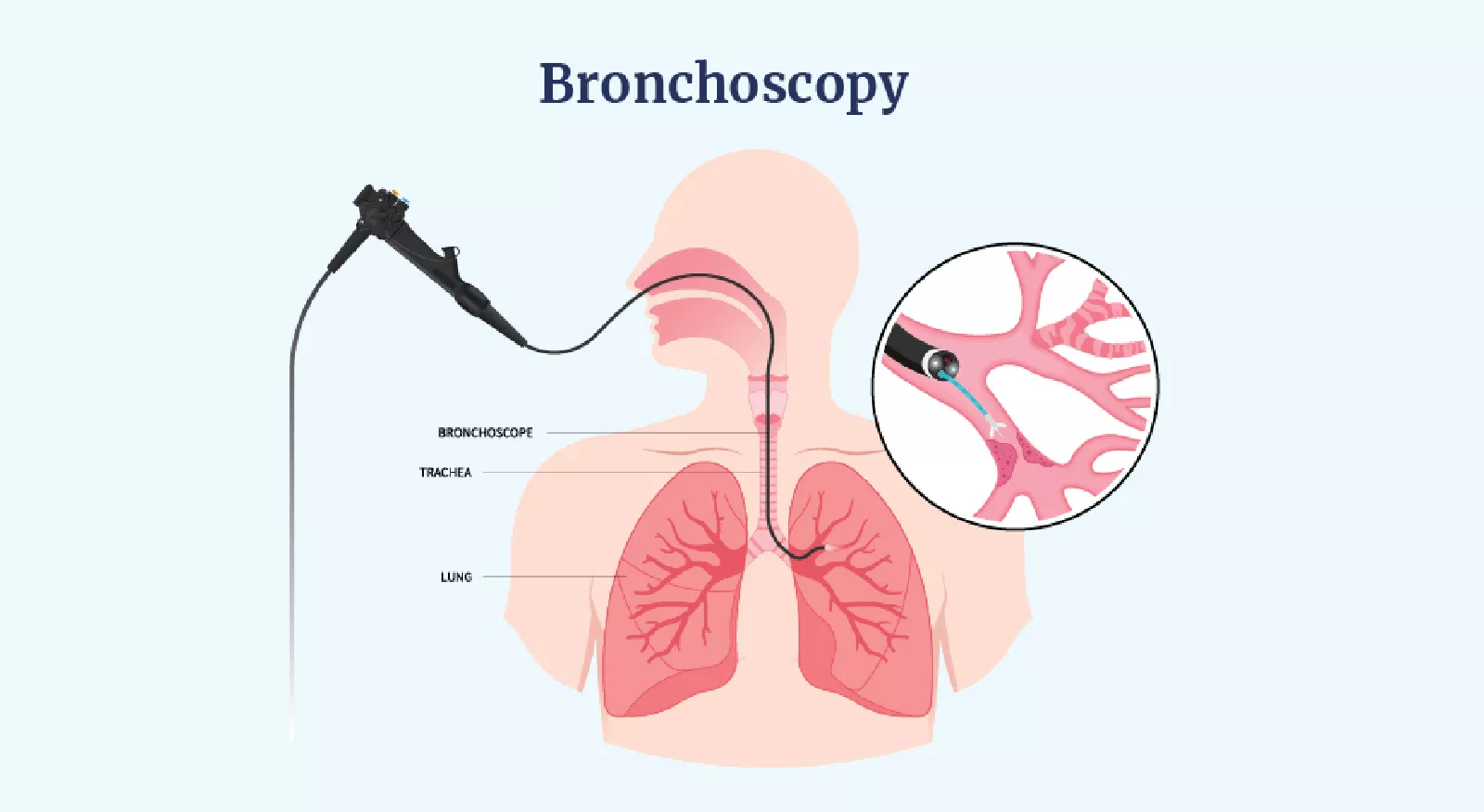
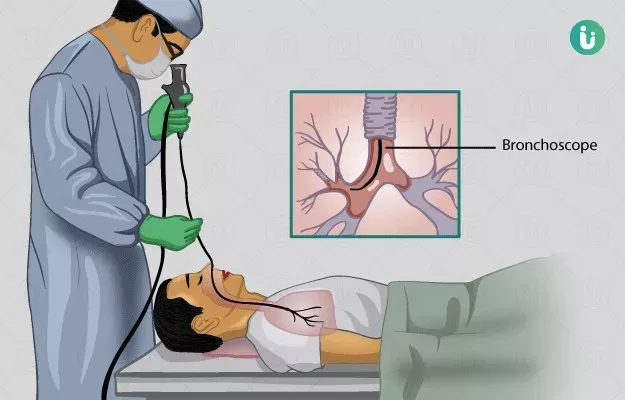
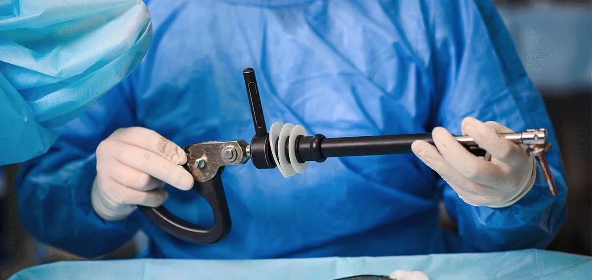
A bronchoscope, a thin flexible tube equipped with a camera, was gently passed through Rahul’s mouth into his airways under mild sedation. This allowed the doctor to directly visualise the right upper lobe lesion seen on the CT scan.
Using real-time camera guidance, the pulmonologist carefully advanced a special biopsy tool through the bronchoscope and collected tissue samples from the lesion. This minimally invasive approach ensured precision, avoiding damage to healthy lung tissue while accurately sampling the suspicious area.
The Biopsy Results: Confirming Cancer
The Biopsy results confirmed the presence of non-small cell lung cancer (NSCLC). Though the news was difficult to process, the early discovery offered a hopeful outlook. However, a critical next step remained—staging the cancer to determine if it had spread to nearby lymph nodes or other parts of the body. To assess this accurately, the pulmonologist recommended a PET-CT scan followed by a specialised procedure called EBUS (Endobronchial Ultrasound).
Step 2: EBUS for Cancer Staging – Why It Was Essential
Why Was EBUS Necessary?
Even though Rahul’s tumor was localised, cancer can spread to the regional lymph nodes in the chest (mediastinal nodes). Knowing whether cancer has spread is crucial for deciding treatment—whether surgery alone is sufficient or if chemotherapy and radiation are required.
How EBUS Was Performed for Rahul
An EBUS bronchoscope, equipped with a miniature ultrasound probe, was inserted into Rahul’s airways under sedation. The ultrasound probe allowed the pulmonologist to see deeper structures, including the lymph nodes surrounding his breathing tubes, which are not visible with regular bronchoscopy.
During the procedure:
• Fine needle aspiration (FNA) was performed to collect small tissue samples from the nearby lymph nodes for microscopic examination.
Step 3: Clear Results and Early Treatment
The results from Rahul’s EBUS biopsy confirmed no distant metastasis. His cancer was staged as early-stage localised lung cancer, meaning it was still confined to the right upper lobe without any spread.
Timely Surgery and a New Lease on Life - Because of the early diagnosis and precise staging using bronchoscopy and EBUS, Rahul underwent a successful surgical resection (removal of the affected part of the lung). Since the tumor was caught at a very early stage.
Today, Rahul remains cancer-free and continues his active lifestyle, grateful for the role that early intervention and advanced diagnostic tools played in his recovery.
