Introduction
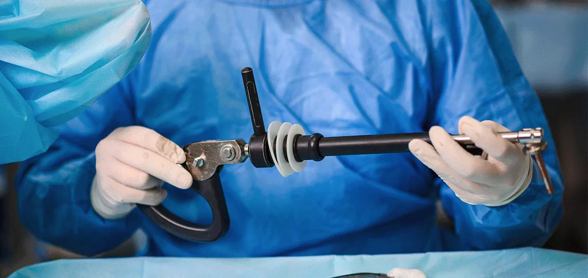
Pleuroscopy involves the passage of an endoscope through the chest wall and helps in the direct visualisation of the parietal and visceral pleura. This is a tool used for both diagnostic and therapeutic management of pleural effusion. Even though VATS (Video Assisted Thoracoscopic Surgery ) is considered to be the definitive diagnostic tool for the diagnosis of undiagnosed pleural effusion, given the increased intra and post-op complications and morbidity medical thoracoscopy/pleuroscopy is increasingly used nowadays for the diagnosis of undiagnosed non-loculated pleural effusion. Patients looking for Pulmonologists In JP Nagar Bangalore can benefit from this minimally invasive approach. Here in this article, we will be discussing the efficiency and usage of pleuroscopy in detail.
Case history
A 47-year-old female, known diabetic and hypothyroid came with complaints of dry cough and breathlessness for 6 months. She also had a history of weight loss of about 20 kgs in 1 year. No history of fever/chest pain. She was diagnosed to have Bilateral pleural effusion (left > right ). She underwent therapeutic thoracentesis initially which revealed an exudative pleural effusion, with low ADA (8.2), lymphocytic predominant, Xpert MTB and AFB smear were negative and cytology was negative for malignant cells. The pleural fluid culture showed no growth. She had recurrent symptoms and a repeat Chest X-ray showed an increase in pleural effusion on the left side for which Intercoastal drainage was done under local anaesthesia. PET CT was done which showed peribronchial vascular interstitial thickening with numerous micro and macro nodules in both lungs suggesting infectious aetiology / lymphangitis carcinomatosis. Few enlarged mediastinal and hilar lymph nodes and prominent bilateral inguinal and distal external iliac lymph nodes. However, in view of recurrent refilling of effusion and drainage, she was referred to the best pulmonologist in Bangalore at Aster RV hospital for further management.
After getting informed consent, pleuroscopy guided pleural biopsy was done under and the sample was sent for culture, Xpert MTB and histopathological examination. Pleural tissue was negative for acid-fast bacilli, Xpert MTB and culture showed no growth. However histopathological examination of the pleural tissue showed features positive for malignancy, features of mucin-secreting adenocarcinoma, primary from lung versus metastasis from other organs such as the gastrointestinal tract or ovary need to be excluded and advised immunohistochemistry for further evaluation.
Discussion
In the United Kingdom, a study was conducted and BTS guidelines were formulated in 2010 which showed that even though traditionally closed blind pleural biopsy was followed as a diagnostic tool for the undiagnosed pleural effusion which increased the diagnosis of malignancy by 7-27% compared to pleural fluid cytology alone, local anaesthetic medical thoracoscopy has increased the diagnostic yield by 92.6 %. This study also compared CT-guided biopsy to the medical thoracoscopy however Ct guided biopsy was not as sensitive as thoracoscopy since not all the pleural nodules are visible on the CT chest. Meta-analysis of 8 studies showed results in which a prior closed blind biopsy was negative, however subsequent thoracoscopy guided biopsy was positive.
But VATS still remains the definitive diagnostic tool for any undiagnosed pleural effusion. But patients should be subjected to VATS only if there is a need for concurrent minimally invasive surgery like tumour resection or excision biopsy, bullectomy, or decortication. This is because of the complexity of the procedure, higher morbidity and higher rates of complications.
A cross-over randomized control trial (COFFEE TRIAL ) was conducted comparing the diagnostic yield of cryobiopsy versus flexible forceps biopsy. In this study FFB and CB was conducted randomly in a 1:1 ratio. The primary outcome was the diagnostic yield of FFB versus CB. The secondary outcomes include sample size, depth, histopathology of the biopsy specimen and the complications such as bleeding during the procedure and the duration of the procedure. This study concluded that even though the sample size was larger, the complications and diagnostic yields are similar in both procedures.
Medical thoracoscopy biopsy can also be used in the diagnosis of diffuse parenchymal lung disease, pulmonary infiltrates or peripheral diseases of unknown cause. Even though bronchoscopy-guided biopsy/cryobiopsy are usually used as a diagnostic tool, a pleuroscopy-guided biopsy can also be used in the peripherally located lesion given the increased sample size, and biopsy can be taken under direct visualisation.
Therapeutic indications
Pleuroscopy also has multiple therapeutic indications. It can be used to remove adhesions and loculations in long-standing pleural effusions. It can be used as a tool for pleurodesis using sclerosant under direct visualisation. Also, Pleural indwelling catheters are being increasingly used for mechanical pleurodesis of the pleura.
A prospective 12-month multicentre trial was conducted in Australia comparing the options of sclerosant-induced pleurodesis and pleural indwelling catheter in patients with malignant pleural effusion. The endpoints of this study were the duration of hospital stay from procedure to death and complication rates like infection, protein depletion and infection was also monitored. According to this study, the patients treated with indwelling pleural catheters had fewer days in the hospital compared to those who received sclerosant-based pleurodesis, also these patients required lesser additional pleural procedures compared to the other group. However, there was no difference in the rate of infection, loss of protein or albumin in both these groups. Patients looking for expert medical care can consult a Pulmonology Hospital in JP Nagar Bangalore for timely and accurate management.
Uncommon applications
Medical thoracoscopy is rarely used in blebectomy , pleural abrasions, foreign body removal or symphatectomy, however, VATS has been proven to be the definitive diagnostic tool for all the above procedures. But given the higher complication rates, longer hospital stays and the requirement of deeper anaesthesia medical thoracoscopy can be tried but with lower success rates compared to VATS.
Conclusion
From the above case, it is evident that pleuroscopy has proved to be a very useful tool in the diagnosis of long-standing undiagnosed pleural effusion. Instead of trying closed pleural biopsy which has a lower diagnostic yield, the patient can be offered pleuroscopy by which we can visualise the pleura completely, take a biopsy from the abnormal areas and also drain the effusion. Removal of adhesions , placement of indwelling pleural catheter , sclerosant pleurodesis can all be done under direct visualisation . Hence pleural effusions in which diagnosis cannot be attained through ultrasound-guided tapping medical thoracoscopy and biopsy should be done for early diagnosis and hence early treatment for the underlying disease process.


